

Functional magnetic resonance imaging
What is Functional magnetic resonance imaging (fMRI)?
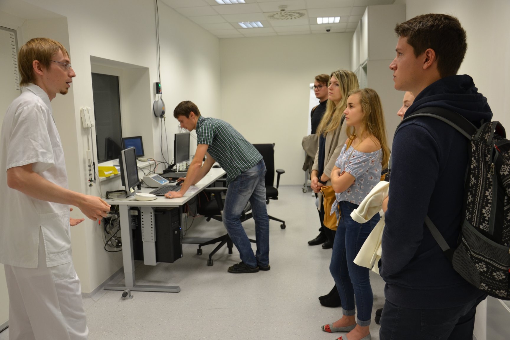
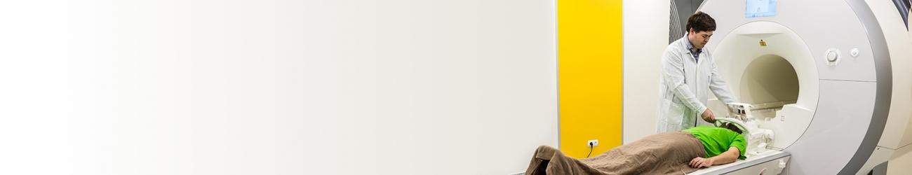
Functional magnetic resonance imaging (fMRI) is a modern neuroimaging method, which is mapping functional areas of the brain activated during a task or stimulation. fMRI began to develop especially in the last decade of the 20th century and greatly enriched knowledge in the field of cognitive neuroscience and clinical neurophysiology. Mapping is performed either on the basis of a change in blood flow to a given area (perfusion) or on the basis of a change in blood oxygenation (the so-called BOLD effect). BOLD fMRI (named after the use of the BOLD effect) is the most common way today and has almost become synonymous with the more general name fMRI. Thus, the method makes it possible, based on changes in blood oxygenation and local blood flow, to indirectly detect those parts of the cerebral cortex that are involved in performing a cognitive, motor, or other task performed by the measured subject. Functional MRI finds application mainly in neurophysiological research. The use in clinical practice is still limited (especially the responsible account of the examined person is needed), however, signs of change are beginning to appear. In many workplaces, this method is already used as an additional examination, eg before a neurosurgical intervention. Suitable clinical applications include the location of speech or motor centers.
Functional brain imaging techniques
Functional brain imaging (or also functional mapping) can be defined in various ways. If we consider a broader definition, then it includes the whole range of techniques in which physiological changes are associated with brain activity. Probably the best method would be to map the activity of individual neurons, which, however, is an almost unsolvable task for imaging methods. Therefore, functional mapping methods rely on the activity of large populations of neurons. Fortunately, this is also an approach with also highly informative value, as the individual neurons do not work independently, but associate into certain functional areas. Thus, despite the very small dimensions of neurons, we are able to map brain functions even using a spatial resolution of units of millimeters or better.
Currently, a number of methods are used for functional mapping based on different principles and using different measured quantities. E.g. EEG (electroencephalograph) senses changes in electrical potentials on the surface of the head, MEG (magnetoencephalography) similarly detects changes in the magnetic field. PET (positron emission tomography) changes in metabolism or blood flow. Relatively young is a method called functional magnetic resonance imaging (fMR, fMRI), which uses an MR tomograph for functional mapping. fMRI can use two methods of indirect mapping of neuronal activity. The first method uses a local increase in blood flow, so-called perfusion, at the site of neuronal activity. The second method uses a change in the ratio of oxygenated and non-oxygenated blood at the site of neuronal activity, which is referred to as the blood oxygenation level-dependent effect. The methods are then called BOLD fMRI and perfusion fMRI. For self-functional imaging, fMRI uses MR images weighted to capture the effects described above.
These methods differ from each other in spatial and temporal resolution. The highest temporal resolution, but at the same time low spatial resolution, is provided by EEG and MEG. The disadvantage of PET is in particular the exposure of the examined person to radioactive radiation and poor time resolution. fMRI is non-invasive, with relatively high spatial resolution and acceptable temporal resolution. It may seem like an ideal method, but it also has its negatives, such as contraindications for examinations in people with a pacemaker, time and economic demands, etc. Of course, these methods are not a complete list but include the most used options for functional brain imaging.